NEWS DETAIL
Seek the TRUTH of Life
Cobio Urine Sediment Smart Technology
Urine analysis are frequently performed on both hospitalized patients and outpatients to screen for diseases of the kidney and urinary tract. The traditional strategy recommended by the European Urinalysis Guidelines involves a two-step procedure, in which semi-quantitative dipstick tests are used in the first step to exclude urine samples without hemoglobin, leukocyte esterase activity, nitrite and protein from further analysis. In a second step, urine samples with indications for erythrocyturia, leukocyturia, bacteriuria or proteinuria are subjected to centrifugation to sediment components for further analysis by microscopy. The microscopic examination of urine for its cellular constituents is a time-consuming procedure requiring expert knowledge and is influenced by preanalytical and analytical processes such as centrifugation and discrepant interpretation of urine sediment structures by technicians, respectively. These limitations cause imprecision and inaccuracy. Standardization of urine analysis has been proposed in the 1995 National Committee for Clinical Laboratory standards (NCCLS) international guidelines and the 2000 European Urinalysis Guidelines.
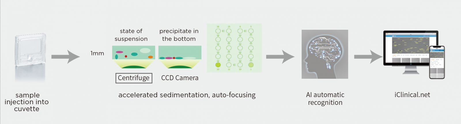
An automated urine sediment analyzer is possibly helpful for standardization and quantification. Cobio Smart Healthcare Technology developed Cobio Systems for complete automation of manual urine microscopy testing. The Cobio S50/S80 system automatically mixes and aspirates samples from tubes. The sample is then injected into a cuvette which is the only consumable needed for the testing. This cuvette then automatically moves to build in centrifuge unit and spun at 2000rpm for 10 seconds. The cuvette then is moved to the microscopy unit where high-definition microscope camera capture the images. Particle images isolated from each of the 15-20 captured frames are identified and labelled using AI deep learning algorithm into 12 categories based on their size, shape, contrast and structure. The high-definition images can be displayed on the screen for verification and manual editing, if necessary. The build in touch screen makes the system easier to handle or user friendly. Results for microscopic particles can be reported as particles per field of view (per high-power field; HPF) or per microliter. The future is now.